The detail visibility is about ½ of image unsharpness (in a "normal" image with "standard noise").
Image Unsharpness (U
Im) depends on
- Focal Spot size (FS)
- Spatial Detector Resolution (SR
b)
- geometric magnification (v) [hint: "v" for magnification is taken out of ASTM E1000; in German language
magnificaiton is
Vergroesserung]
This three factors are in the formula for image unsharpness in ASTM E1000:
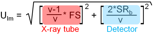
Formula
[1]The two factors in the square root contains the tube and the detector and the magnification. With given tube and detector the magnification is the variable which can be used to optimize the detail visibility or minimum defect discernable. In E1000 is a graph in which the detail visibility is normalized to the detector unsharpness U
f or the visibility of the system with no magnification (v=1) - as used with films before:
The different lines in the graph show different relation ships between focal spot size (phi) and detector unsharpness U
f. With a large focal spot - in the graph [(phi)/U
f=8] already a small magnification will lead to a higher unsharpness as the detector unsharpness. As the film is really sharp but need high dose, large focal spots are used for film exposure and the film is normally in close contact with the object to be inspected. With a small focal spot the lines decrease and details smaller than the detector resolution will be visible. µ-focus systems use high magnifications with very small focal spots for electronic inspection where resolutions in µm range are required.
If we look at the two extremas, we get a better feeling for what is in the graph.
1. No magnification (v=1) [object directly on detector like with film]. In Formula [1] the factor with the focalspot size FS (or [phi]) is Zero as at v=1 the quotient in front of FS is zero.
Finally the Image Unsharpness of the system is the detector unsharpness or two times the spatial resolution of the detector (SR
b).
2. Very high magnification (v>>100) [object very close to tube head]. In Formula [1] the factor with the detector unsharpness decreases with the magnification and it becomes infinitesimal small. The quotient in front of the FS mutates to 1.
Finally with very high magnifications the Image Unsharpness of the system is the focal spot size FS.
Between this two extrema all X-ray applications are settled. Having in mind that the
it is obvious the small focal spots give only low dose.


 ).
).